AI-Based Classification of Mandibular Distal Root Canal Curvature: Using Deep Learning on Panoramic Radiographs
AI-Based Classification of Mandibular Distal Root Canal Curvature
DOI:
https://doi.org/10.63841/iue24519Keywords:
Deep Learning, Endodontic Diagnosis, Root Canal Curvature, Schneider’s Method, Panoramic Radiographs (OPG)Abstract
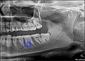
Root canal morphology, particularly the curvature of the root canals, plays a crucial role in the efficacy of endodontic diagnosis and treatment planning. Yet, assessing this morphology typically relies on manual annotation and interpretation of dental radiographs, which can be time-consuming and prone to human error. These challenges are especially pronounced in low-resource settings such as Kurdistan, the northern part of Iraq, where access to precision imaging tools is limited. This study proposes a novel AI-based framework for classifying mandibular distal root canal curvature. Furthermore, it introduces a semi-automated annotation pipeline that integrates Schneider’s method with AutoCAD, facilitating the labeling of root canal curvatures for deep learning applications.
Initially, a total number of 11,490 OPG images were gathered from public and private clinics. After undergoing rigorous preprocessing, 1,166 OPG images were selected and categorized into three classes: straight, moderate, and severe, based on Schneider’s method for root canal curvature classification. Three pre-trained convolutional neural network models — ResNet-101, DenseNet-121, and EfficientNet-B0 — were fine-tuned on the collected OPGs. ResNet-101 was evaluated using a fivefold cross-validation test, while DenseNet-121 and EfficientNet-B0 were fine-tuned using a two-phase transfer learning strategy. In this approach, the initial feature extractor was first frozen, followed by fine-tuning of the whole model. To address the limited sample size, data augmentation techniques were also employed to mitigate overfitting.
Among those three models, ResNet-101 achieved the highest classification accuracy, 0.907, and an F1-score of 0.907, demonstrating superior ability to capture anatomical features. These findings underscore the effectiveness and promising role of deep learning in supporting dental diagnosis, particularly in endodontics. The proposed AI framework, combined with a semi-automated annotation approach, has the potential to improve the scalability and accessibility of root canal morphology assessment even in low-resource clinical settings.
Downloads
References
R. Fuentes, C. Farfán, N. Astete, P. Navarro, and A. Arias, “Distal root curvatures in mandibular molars: Analysis using digital panoramic X-rays,” Folia Morphologica (Poland), vol. 77, no. 1, pp. 131–137, 2018, doi: 10.5603/FM.a2017.0066.
F. J. Vertucci, “Root canal anatomy of the human permanent teeth,” Oral Surgery, Oral Medicine, Oral Pathology, vol. 58, no. 5, pp. 589–599, 1984, doi: 10.1016/0030-4220(84)90085-9.
S. W. Schneider, “A comparison of canal preparations in straight and curved root canals,” Oral Surgery, Oral Medicine, Oral Pathology, vol. 32, no. 2, pp. 271–275, 1971, doi: 10.1016/0030-4220(71)90230-1.
C. Lucena, E. M. Souza, G. C. Voinea, R. Pulgar, M. J. Valderrama, and G. De-Deus, “A quality assessment of randomized controlled trial reports in endodontics,” Int Endod J, vol. 50, no. 3, pp. 237–250, 2017, doi: 10.1111/iej.12626.
M. H. Malur and A. Chandra, “Curvature height and distance of MB canal of mandibular molar with Schneider angle and its comparison with canal access angle,” J Oral Biol Craniofac Res, vol. 8, no. 3, pp. 212–216, 2018, doi: 10.1016/j.jobcr.2017.07.002.
P. Balani, F. Niazi, and H. Rashid, “A brief review of the methods used to determine the curvature of root canals,” Journal of Restorative Dentistry, vol. 3, no. 3, p. 57, 2015, doi: 10.4103/2321-4619.168733.
T. Hiraiwa et al., “A deep-learning artificial intelligence system for assessment of root morphology of the mandibular first molar on panoramic radiography,” Dentomaxillofacial Radiology, vol. 48, no. 3, pp. 1–7, 2019, doi: 10.1259/dmfr.20180218.
S. Asgary, “Artificial Intelligence in Endodontics: A Scoping Review,” Iran Endod J, vol. 19, no. 2, pp. 85–98, 2024, doi: 10.22037/iej.v19i2.44842.
E. G. Ha, K. J. Jeon, H. Choi, C. Lee, Y. J. Choi, and S. S. Han, “Automatic diagnosis of retention pseudocyst in the maxillary sinus on panoramic radiographs using a convolutional neural network algorithm,” Sci Rep, vol. 13, no. 1, pp. 1–7, 2023, doi: 10.1038/s41598-023-29890-5.
V. Roda-Casanova, A. Pérez-González, Á. Zubizarreta-Macho, and V. Faus-Matoses, “Fatigue analysis of NiTi rotary endodontic files through finite element simulation: Effect of root canal geometry on fatigue life,” J Clin Med, vol. 10, no. 23, 2021, doi: 10.3390/jcm10235692.
S. J. Jeon et al., “Deep-learning for predicting C-shaped canals in mandibular second molars on panoramic radiographs,” Dentomaxillofacial Radiology, vol. 50, no. 5, pp. 1–6, 2021, doi: 10.1259/dmfr.20200513.
L. Zhang et al., “A lightweight convolutional neural network model with receptive field block for C-shaped root canal detection in mandibular second molars,” Sci Rep, vol. 12, no. 1, pp. 1–11, 2022, doi: 10.1038/s41598-022-20411-4.
B. Çelik and M. E. Çelik, “Root Dilaceration Using Deep Learning: A Diagnostic Approach,” Applied Sciences (Switzerland), vol. 13, no. 14, 2023, doi: 10.3390/app13148260.
U. Rashid et al., “A hybrid mask RCNN-based tool to localize dental cavities from real-time mixed photographic images,” PeerJ Comput Sci, vol. 8, pp. 1–24, 2022, doi: 10.7717/PEERJ-CS.888.
R. C. Hartmann, M. Fensterseifer, O. A. Peters, J. A. P. de Figueiredo, M. S. Gomes, and G. Rossi-Fedele, “Methods for measurement of root canal curvature: a systematic and critical review,” Int Endod J, vol. 52, no. 2, pp. 169–180, 2019, doi: 10.1111/iej.12996.
A. K. Navas, J.M., Doranala, S., Khushnud, A., Sinha, J., Jadhav, A.A., Gudapati, S. and Syed, “Methodology Used in the Study,” J Pharm Bioallied Sci, 2022, doi: 10.4103/jpbs.jpbs_714_21.
M. T. Z. Aung et al., “Deep learning-based automatic segmentation of the mandibular canal on panoramic radiographs: A multi-device study,” Imaging Sci Dent, vol. 54, no. 1, pp. 81–91, 2024, doi: 10.5624/isd.20230245.
H. Nekkanti, S. Enuganti, J. S. S. Avula, P. Kakarla, S. K. Siva, and D. N. Gavarraju, “Assessment of Biomechanical Preparation Influence on Various Root Canal Curvatures,” Int J Clin Pediatr Dent, vol. 17, no. 2, pp. 10–15, 2024, doi: 10.5005/jp-journals-10005-2760.
K. He, X. Zhang, S. Ren, and J. Sun, “Deep residual learning for image recognition,” Proceedings of the IEEE Computer Society Conference on Computer Vision and Pattern Recognition, vol. 2016-Decem, pp. 770–778, 2016, doi: 10.1109/CVPR.2016.90.
M. Tan and Q. V. Le, “EfficientNet: Rethinking model scaling for convolutional neural networks,” 36th International Conference on Machine Learning, ICML 2019, vol. 2019-June, pp. 10691–10700, 2019.
G. Huang, Z. Liu, G. Pleiss, L. Van Der Maaten, and K. Q. Weinberger, “Convolutional Networks with Dense Connectivity,” IEEE Trans Pattern Anal Mach Intell, vol. 44, no. 12, pp. 8704–8716, 2022, doi: 10.1109/TPAMI.2019.2918284.
A. Khan et al., “Human gait recognition using deep learning and improved ant colony optimization,” Computers, Materials and Continua, vol. 70, no. 2, pp. 2113–2130, 2022, doi: 10.32604/cmc.2022.018270.
A. Hussain, S. Ul Amin, M. Fayaz, and S. Seo, “An Efficient and Robust Hand Gesture Recognition System of Sign Language Employing Finetuned Inception-V3 and Efficientnet-B0 Network,” Computer Systems Science and Engineering, vol. 46, no. 3, pp. 3509–3525, 2023, doi: 10.32604/csse.2023.037258.
R. Chetan, D. V Ashoka, and A. Prakash, “HybridTransferNet: Advancing Soil Image Classiication through Comprehensive Evaluation of Hybrid Transfer Learning HybridTransferNet: Advancing Soil Image Classification through Comprehensive Evaluation of Hybrid Transfer Learning,” 2023, [Online]. Available: https://doi.org/10.21203/rs.3.rs-3032907/v1
Downloads
Published
Issue
Section
License
Copyright (c) 2025 Academic Journal of International University of Erbil

This work is licensed under a Creative Commons Attribution-NonCommercial-NoDerivatives 4.0 International License.